
1185 W Carmel Dr. #D2, Carmel, IN 46032

Understanding Bone Loss and Root Canal Success: A Complete Patient Guide

Persistent tooth pain, severe temperature sensitivity, or localized jaw discomfort can significantly disrupt your daily routine, whether you are enjoying a weekend morning at the Carmel Farmers Market or biking along the Monon Trail. When internal tooth decay or pulp infection progresses, it frequently triggers inflammation that reaches the surrounding jawbone. Many patients facing this situation worry whether pre-existing or infection-induced bone loss will permanently compromise their treatment outcome.
At Alvarez Family Dentistry, Dr. Keith Alvarez provides detailed clinical evaluations to determine the exact cause of tissue deterioration and restore your oral health. If you are experiencing persistent dental discomfort while enjoying local spots around town, receiving timely root canal therapy in Carmel can prevent infection from spreading into the surrounding alveolar bone. Book an appointment today to protect your natural smile.
How Alveolar Bone Loss Interacts with Root Canal Therapy
Bone loss does not automatically cause root canal failure, but the underlying cause and extent of the bone loss directly dictate long-term prognosis. When bone destruction stems entirely from an infected tooth root, successful endodontic cleaning typically allows the surrounding jawbone to regenerate fully over time. However, if the bone loss is caused by advanced generalized gum disease, endodontic treatment alone cannot rebuild lost structural support.
To understand how bone loss affects treatment outcomes, it is crucial to distinguish between endodontic bone loss and periodontal bone loss.
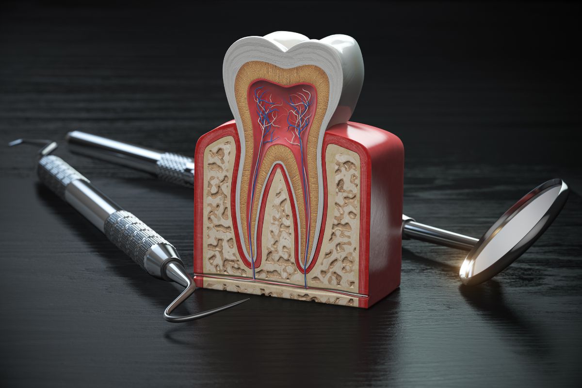
- Endodontic Bone Loss: Occurs when bacteria inside the tooth’s canal system travel through the apical foramen (the tip of the root) into the surrounding bone. This localized response forms an apical lesion or abscess. Once the root canal system is thoroughly disinfected and sealed, the stimulus for bone destruction is removed, allowing osteoblasts (bone-forming cells) to repair the socket.
- Periodontal Bone Loss: Results from chronic marginal gum disease (periodontitis). Bacteria erode the attachment fibers and alveolar crest from the gumline downward. This form of bone destruction is systemic to the periodontal pocket rather than isolated to the root tip.
According to research published by the Centers for Disease Control and Prevention (CDC), approximately 47.2% of adults aged 30 and older in the United States suffer from some form of periodontal disease. When periodontitis and pulp necrosis occur simultaneously on the same tooth, clinicians diagnose a combined “endo-perio” lesion. Treating these cases requires a coordinated approach to address both internal canal infection and external gum attachment loss.
Endodontic vs. Periodontal Bone Loss: Diagnostic Comparison
Accurate diagnosis is the cornerstone of successful endodontic therapy when jawbone density is diminished. The source of bone loss dictates whether a tooth requires root canal treatment, periodontal therapy, or a combination of both.
| Diagnostic Factor | Endodontic Bone Loss | Periodontal Bone Loss | Combined Endo-Perio Lesion |
|---|---|---|---|
| Primary Cause | Internal nerve infection or pulp necrosis | Plaque accumulation and chronic gum disease | Dual infection of pulp tissue and deep periodontal pockets |
| Location of Bone Defect | Isolated around the root apex (tip) | Horizontal or vertical loss around the cervical neck | Continuous defect extending from root tip to gum margin |
| Tooth Vitality | Non-vital (dead pulp tissue) | Typically vital (unless secondary infection occurs) | Non-vital |
| Prognosis After Root Canal | Excellent (high rate of bone regeneration) | Guarded (requires ongoing periodontal management) | Variable (depends on remaining periodontal attachment) |
Can a Tooth with Significant Bone Loss Still Be Saved?
A tooth with bone loss can often be saved if the remaining structural support is stable and the root canal system can be completely disinfected. The overall success rate depends heavily on periodontal pocket depth, tooth mobility, and the height of the remaining alveolar bone. Determining whether a compromised tooth can withstand functional chewing forces requires a precise clinical evaluation, making specialized root canal therapy in Carmel a vital intervention for preserving natural bite mechanics.
When assessing whether a compromised tooth is viable for endodontic preservation, clinicians evaluate four main structural criteria:
- Crown-to-Root Ratio: The proportion of tooth structure above the bone compared to the root embedded within the bone. A favorable ratio ensures the tooth can withstand everyday biting forces without fracturing.
- Mobility Grade: Excessive horizontal or vertical movement indicates that periodontal ligament support has been severely compromised, reducing predictable long-term retention.
- Root Anatomy & Curvature: Complex, curved, or calcified canals require specialized endodontic instruments to ensure complete eradication of bacterial biofilms.
- Biological Width: The vertical distance required for healthy gum tissue attachment above the alveolar crest must remain intact to prevent chronic inflammation.
Advanced Healing Protocols and Bone Regeneration Techniques
Bone tissue possesses a remarkable capacity for self-healing once root canal disinfection is complete. Modern endodontic protocols utilize bioceramic sealers and 3D digital imaging to create a sterile internal environment, enabling trabecular bone to fill in previous areas of destruction over 6 to 12 months.
In complex scenarios involving extensive bone destruction, additional clinical therapies may be integrated:
- Bioceramic Root Fillings: Highly biocompatible materials that interact with tissue fluids to form hydroxyapatite, promoting active bone apposition at the root apex.
- Targeted Periodontal Grafting: Placing synthetic or natural bone graft material alongside guided tissue regeneration membranes to reconstruct periodontal walls.
- Crown Lengthening or Restorative Reconstruction: Adjusting hard and soft tissue margins to allow secure placement of a protective dental crown without impinging on periodontal health.
Preserving your natural teeth allows you to continue enjoying local dining experiences from casual lunches near Clay Terrace to dinner with family at local spots like Bub’s Burgers without worrying about bite discomfort or compromised chewing ability.
Long-Term Maintenance and Success Factors
Achieving lasting success after endodontic treatment on a tooth with bone loss requires continuous post-treatment care and proactive maintenance. Eliminating internal infection creates the foundation for recovery, but maintaining surrounding bone height depends on daily oral hygiene and routine clinical monitoring.
- Prompt Restorative Coverage: Placing a full-coverage crown immediately following root canal therapy seals the internal filling from salivary contamination and prevents structural fractures.
- Rigorous Plaque Control: Brushing, flossing, and using antimicrobial rinses prevent marginal bacteria from invading deep periodontal pockets.
- Routine Radiographic Monitoring: Follow-up X-rays at 6-month and 12-month intervals allow clinicians to verify active bone re-mineralization around the root apex.
When you visit our dental office, our clinical team conducts comprehensive 3D digital imaging to assess your trabecular bone density and periodontal health. Providing comprehensive root canal therapy in Carmel enables us to deliver precise care tailored to your unique anatomical needs.
Summary & Next Steps
Bone loss does not spell the end for an infected tooth. While severe periodontal destruction requires careful co-management, isolated endodontic bone loss responds predictably to thorough canal disinfection and sealing. Taking prompt action at the first sign of pain or swelling is the most effective way to prevent localized bone destruction from escalating. Contact Alvarez Family Dentistry today to book an appointment for a comprehensive clinical assessment and personalized care plan.
Frequently Asked Questions
1. Does bone loss lower the success rate of a root canal?
Isolated bone loss caused by tooth infection does not lower success rates once properly disinfected. However, severe pre-existing periodontal bone loss can reduce overall long-term tooth stability.
2. Can jawbone grow back after a root canal?
Yes, bone destroyed by an apical infection routinely regenerates after root canal treatment removes the bacterial source. Complete trabecular bone re-mineralization typically occurs over 6 to 12 months.
3. How do dentists treat a root canal infection with bone loss?
Dentists thoroughly clean, disinfect, and seal the root canals to eradicate internal bacteria. Removing the infectious stimulus allows the body’s natural osteoblasts to rebuild healthy bone around the root tip.
4. How can I tell if my bone loss is from a tooth infection or gum disease?
Endodontic bone loss causes localized pain around a non-vital root tip, visible on X-rays. Periodontal bone loss causes generalized gum recession, deep pockets, and bleeding around multiple vital teeth.
5. Will I need a bone graft during a root canal?
Most standard root canals do not require bone grafting because disinfection prompts natural bone healing. Severe periodontal defects or surgical apicoectomies, however, may benefit from supplemental bone grafting.
6. Is a root canal better than pulling a tooth with bone loss?
Preserving your natural root structure helps maintain jawbone volume and prevents adjacent teeth from shifting. Extraction is reserved only for teeth with non-restorable fractures or severe structural mobility.
7. How long does it take for bone to heal after root canal treatment?
Initial tissue healing begins within a few weeks following successful endodontic disinfection and sealing. Complete bone density restoration is monitored via follow-up X-rays over 6 to 12 months.